Nonlactating Mammary Gland


Lactating mammary gland from a guinea pig
The mammary gland consists of 15–25 separate tubuloalveolar glands in the lobules of the mammary gland, which are separated by connective tissue and adipose tissue. Their alveoli are fully developed only in the course of pregnancy and lactation. Alveoli are rarely present in quiescent glands that do not lactate, especially in nulliparous women. This figure shows the central secretory duct (sinus lactiferi) 1 . Tubuli with blind ends and incompletely developed ducts (ductus lactiferi) branch from it. Note the loose sheath of cellular connective tissue 2 . It is clearly distinguished from the coarse fibrous connective tissue stroma 3 . Quiescent, nonlactating human mammary gland.
Figure 591

Info:
1. Sinus lactiferi
2. Connective tissue sheath
3. Coarse fibrous collagenous connective tissue
Stain: alum hematoxylin-eosin; magnification: × 80
2. Connective tissue sheath
3. Coarse fibrous collagenous connective tissue
Stain: alum hematoxylin-eosin; magnification: × 80
Lactating Mammary Gland
The secretory ducts of the mammary gland start to sprout during pregnancy. Alveoli and lobules form. The connective tissue recedes and the gland parenchyma increases. At the height of lactation (the figure), differently shaped alveoli are found in close proximity of each other. They are separated by delicate connective tissue fibers. The gland epithelium 1 has different heights, dependent on its secretory state. Lactating mammary gland. Secretory products are visible in some of the gland lumina (cf. Fig. 593).
The secretory ducts of the mammary gland start to sprout during pregnancy. Alveoli and lobules form. The connective tissue recedes and the gland parenchyma increases. At the height of lactation (the figure), differently shaped alveoli are found in close proximity of each other. They are separated by delicate connective tissue fibers. The gland epithelium 1 has different heights, dependent on its secretory state. Lactating mammary gland. Secretory products are visible in some of the gland lumina (cf. Fig. 593).
Figure 592

Info :
1. Alveoli
2. Connective tissue septa
Stain: azan; magnification: × 80
2. Connective tissue septa
Stain: azan; magnification: × 80
In the active, lactating mammary gland, the epithelium of the alveoli features a rich ergastoplasm and apical fat droplets. It shows different heights. The usual preparations display round empty spaces 1 where fat droplets have been (cf. Figs. 594, 595). The apical cell membrane may rupture or, in other places, bulge dome-like into the lumen. These attributes are characteristic of apocrine glands (see Fig.133). There are patches with secretory product 2 and occasionally sloughed-off epithelial cells (upper figure). Elaborately branchedmyoepithelial cells 3 form an incomplete layer around the alveolar wall.
Figure 593

Info :
1. Secretory product in gland cells (vacuoles)
2. Secretory product
3. Myoepithelial cellsStain: alum hematoxylin-eosin; magnification: × 160
2. Secretory product
3. Myoepithelial cellsStain: alum hematoxylin-eosin; magnification: × 160
The fat droplets of the milk are blackened with osmium tetroxide in this section of a lactating mammary gland. They correspond to the vacuoles, which are seen in Fig. 593. Note that small droplets combine to larger spheres in some places. The gland cells and the connective tissue are stained yellowish brown.
Figure 594

Stain: osmium tetroxide; magnification: × 160
The lactating mammary gland concomitantly synthesizes several different substances, which are released into the alveoli. During periods of lactation, the secretory cells produce fat droplets (see Fig.594). They combine to larger droplets and are moved to the apical cell region. Finally, they become part of vesicular formations at the plasmalemma (apical protrusions) and are pinched off as milk droplets.
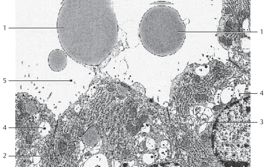
The figure displays two membrane encased fat droplets 1 in the alveolar lumen. The large central, membrane encased fat droplet is still connected to the plasmalemma. It will obviously be pinched off shortly. At the same time, the ergastoplasm 2 of the gland cells in conjunction with the Golgi apparatus biosynthesize proteins, in particular casein and α-lactalbumin and packaged as secretory vesicles. The small casein granules 4 have a dense structure. They are osmiophil and can therefore easily be found. Casein granules fuse with the plasmalemma and are released by exocytosis. Four gland cells with many protein vesicles 4 are visible in this section. The gland cells have an elaborate endoplasmic reticulum 2 (ergastoplasm), Golgi complexes 3 and mitochondria. Sporadically, casein granules are already present in the lumen of the alveoli 5 .
The figure displays two membrane encased fat droplets 1 in the alveolar lumen. The large central, membrane encased fat droplet is still connected to the plasmalemma. It will obviously be pinched off shortly. At the same time, the ergastoplasm 2 of the gland cells in conjunction with the Golgi apparatus biosynthesize proteins, in particular casein and α-lactalbumin and packaged as secretory vesicles. The small casein granules 4 have a dense structure. They are osmiophil and can therefore easily be found. Casein granules fuse with the plasmalemma and are released by exocytosis. Four gland cells with many protein vesicles 4 are visible in this section. The gland cells have an elaborate endoplasmic reticulum 2 (ergastoplasm), Golgi complexes 3 and mitochondria. Sporadically, casein granules are already present in the lumen of the alveoli 5 .
Lactating mammary gland from a guinea pig
Figure 595
Info :
1. Membrane-encased fat droplet
2. Ergastoplasm
3. Golgi apparatus
4. Vacuole with casein granule
5. Casein granule in the alveolar lumen
Electron microscopy; magnification: × 6000
2. Ergastoplasm
3. Golgi apparatus
4. Vacuole with casein granule
5. Casein granule in the alveolar lumen
Electron microscopy; magnification: × 6000
Reference :
Kuehnel, Color Atlas of Cytology, Histology, and Microscopic Anatomy, 4th ed. 2003.