Rabu, 29 Juni 2011

Warming Up

When The Doctor was preparing a drug that would be injected to the patient, suddenly, the patient who was lying in bed screaming out. The Doctor was surprised, so he asked to the patient: 
The Doctor : Well...why are you screaming out ? What is the fear of injections ? 
Patients : No...Sir, I screamed just for warming up. So that, when I'm shot I will not scream anymore... :D 
The Doctor : ?!!?!!?!?!

Selasa, 28 Juni 2011

Cytosol

 At one time, it was believed that the cytoplasm intervening between the discrete organelles and deposits was unstructured. This belief was reinforced by the use of homogenization and centrifugation of the homogenates to yield fractions consisting of recognizable membrane-bound organelles. The final supernatant produced by this process, after the separation of organelles, is called the cytosol. The cytosol constitutes about half the total volume of the cell. Homogenization of cells disrupts a delicate microtrabecular lattice that incorporates filaments of actin, microtubules, intermediate filaments, enzymes, and other soluble constituents into a structured cytosol. The cytosol coordinates the intracellular movements of organelles and provides an explanation for the viscosity of the cytoplasm. Soluble (not membrane-bound) enzymes, such as those of the glycolytic pathway, for example, function more efficiently when organized in a sequence instead of having to rely on random collisions with their substrates. The cytosol provides a framework for this organization. It contains thousands of enzymes that produce building blocks for larger molecules and break down small molecules to liberate energy. All machinery to synthesize proteins (rRNA, mRNA, tRNA, enzymes, and other factors) is contained in the cytosol.

Cell Components & Diseases

Many diseases are related to molecular alterations in specific cell components. In several of these diseases, structural changes can be detected by light or electron microscopy or by cytochemical techniques. Table 2–5 lists some of these diseases and emphasizes the importance of understanding the many cell components in pathobiology.
Table 2–5. Some Human and Animal Diseases Related to Altered Cellular Components.
Cell Component Involved Disease Molecular Defect Morphological Change Clinical Consequence
Mitochondrion Mitochondrial cytopathy Defect of oxidative phosphorylation Increase in size and number of muscle mitochondria High basal metabolism without hyperthyroidism
Microtubule Immotile cilia syndrome Lack of dynein in cilia and flagella Lack of arms of the doublet microtubules Immotile cilia and flagella with male sterility and chronic respiratory infection
Mouse (Acomys) diabetes   Reduction of tubulin in pancreatic cells Reduction of microtubules in cells High blood sugar content (diabetes)
Lysosome Metachromatic leukodystrophy Lack of lysosomal sulfatase Accumulation of lipid (cerebroside) in tissues Motor and mental impairment
Hurler disease Lack of lysosomal -L-iduronidase  Accumulation of dermatan sulfate in tissues Growth and mental retardation
Golgi complex I-cell disease Phosphotransferase deficiency Inclusion-particle storage in several cells Psychomotor retardation, bone abnormalities

References
Afzelius BA, Eliasson R: Flagellar mutants in man: on the heterogeneity of the immotile-cilia syndrome. J Ultrastruct Res 1979;69:43. [PMID: 501788]
Aridor M, Balch WE: Integration of endoplasmic reticulum signaling in health and disease. Nat Med 1999;5:745. [PMID: 10395318]
Barrit GJ: Communication Within Animal Cells. Oxford University Press, 1992.
Becker WM et al: The World of the Cell, 4th ed. Benjamin/Cummings, 2000.
Bretscher MS: The molecules of the cell membrane. Sci Am 1985;253:100. [PMID: 2416050]
Brinkley BR: Microtubule organizing centers. Annu Rev Cell Biol 1985;1:145. [PMID: 3916316]
Brown MS et al: Recycling receptors: the round-trip itinerary of migrant membrane proteins. Cell 1983;32:663. [PMID: 6299572]
Cooper GM: The Cell: A Molecular Approach. ASM Press/Sinauer Associates, Inc., 1997.
DeDuve C: A Guided Tour of the Living Cell. Freeman, 1984.
DeDuve C: Microbodies in the living cell. Sci Am 1983;248:74.
Dustin P: Microtubules, 2nd ed. Springer-Verlag, 1984.
Farquhar MG: Progress in unraveling pathways of Golgi traffic. Annu Rev Cell Biol 1985;1:447. [PMID: 3916320]
Fawcett D: The Cell, 2nd ed. Saunders, 1981.
Krstíc RV: Ultrastructure of the Mammalian Cell. Springer-Verlag, 1979.
Mitchison TJ, Cramer LP: Actin-based cell motility and cell locomotion. Cell 1996;84:371. [PMID: 8608590]
Osborn M, Weber K: Intermediate filaments: cell-type-specific markers in differentiation and pathology. Cell 1982;31:303. [PMID: 6891619]
Pfeffer SR, Rothman JE: Biosynthetic protein transport and sorting in the endoplasmic reticulum. Annu Rev Biochem 1987;56:829. [PMID: 3304148]
Rothman J: The compartmental organization of the Golgi apparatus. Sci Am 1985;253:74. [PMID: 3929377]
Simons K, Ikonen E: How cells handle cholesterol. Science 2000;290:1721. [PMID: 11099405]
Tzagoloff A: Mitochondria. Plenum, 1982.
Weber K, Osborn M: The molecules of the cell matrix. Sci Am 1985;253:110. [PMID: 4071030]

Minggu, 26 Juni 2011

Cytoplasmic Deposits

Cytoplasmic deposits are usually transitory components of the cytoplasm, composed mainly of accumulated metabolites or other substances. The accumulated molecules occur in several forms, one of them being lipid droplets in adipose tissue, adrenal cortex cells, and liver cells (Figure 2–38). Carbohydrate accumulations are also visible in several cells in the form of glycogen. After impregnation with lead salts, glycogen appears as collections of electron-dense particles (Figure 2–39). Proteins are stored in glandular cells as secretory granules or secretory vesicles (Figure 2–29); under stimulation, these proteins are periodically released into the extracellular medium.

Figure 2-38

Section of adrenal gland showing lipid droplets (L) and abundant anomalous mitochondria (M). x19,000.

Figure 2-39

Electron micrograph of a section of a liver cell showing glycogen deposits as accumulations of electron-dense particles (arrows). The dark structures with a dense core are peroxisomes. Mitochondria (M) are also shown. x30,000.

Deposits of colored substances—pigments—are often found in cells (Figure 2–40). They may be synthesized by the cell (eg, in the skin melanocytes) or come from outside the body (eg, carotene). One of the most common pigments is lipofuscin, a yellowish-brown substance present mainly in permanent cells (eg, neurons, cardiac muscle) that increases in quantity with age. Its chemical constitution is complex. It is believed that granules of lipofuscin derive from secondary lysosomes and represent deposits of indigestible substances. A widely distributed pigment, melanin is abundant in the epidermis and in the pigment layer of the retina in the form of dense intracellular membrane-limited granules.

Figure 2-40
Section of amphibian liver shows cells with pigment deposit (PD) in the cytoplasm, a macrophage (M), hepatocytes (H), and a neutrophil leukocyte (N). In this resin-embedded material it is possible to see mitochondria (pale red) and lysosomes (blue) in the cytoplasm of the hepatocytes. Only with resin embedding is it possible to obtain such information. Giemsa stain. Medium magnification.

References
Afzelius BA, Eliasson R: Flagellar mutants in man: on the heterogeneity of the immotile-cilia syndrome. J Ultrastruct Res 1979;69:43. [PMID: 501788]
Aridor M, Balch WE: Integration of endoplasmic reticulum signaling in health and disease. Nat Med 1999;5:745. [PMID: 10395318]
Barrit GJ: Communication Within Animal Cells. Oxford University Press, 1992.
Becker WM et al: The World of the Cell, 4th ed. Benjamin/Cummings, 2000.
Bretscher MS: The molecules of the cell membrane. Sci Am 1985;253:100. [PMID: 2416050]
Brinkley BR: Microtubule organizing centers. Annu Rev Cell Biol 1985;1:145. [PMID: 3916316]
Brown MS et al: Recycling receptors: the round-trip itinerary of migrant membrane proteins. Cell 1983;32:663. [PMID: 6299572]
Cooper GM: The Cell: A Molecular Approach. ASM Press/Sinauer Associates, Inc., 1997.
DeDuve C: A Guided Tour of the Living Cell. Freeman, 1984.
DeDuve C: Microbodies in the living cell. Sci Am 1983;248:74.
Dustin P: Microtubules, 2nd ed. Springer-Verlag, 1984.
Farquhar MG: Progress in unraveling pathways of Golgi traffic. Annu Rev Cell Biol 1985;1:447. [PMID: 3916320]
Fawcett D: The Cell, 2nd ed. Saunders, 1981.
Krstíc RV: Ultrastructure of the Mammalian Cell. Springer-Verlag, 1979.
Mitchison TJ, Cramer LP: Actin-based cell motility and cell locomotion. Cell 1996;84:371. [PMID: 8608590]
Osborn M, Weber K: Intermediate filaments: cell-type-specific markers in differentiation and pathology. Cell 1982;31:303. [PMID: 6891619]
Pfeffer SR, Rothman JE: Biosynthetic protein transport and sorting in the endoplasmic reticulum. Annu Rev Biochem 1987;56:829. [PMID: 3304148]
Rothman J: The compartmental organization of the Golgi apparatus. Sci Am 1985;253:74. [PMID: 3929377]
Simons K, Ikonen E: How cells handle cholesterol. Science 2000;290:1721. [PMID: 11099405]
Tzagoloff A: Mitochondria. Plenum, 1982.
Weber K, Osborn M: The molecules of the cell matrix. Sci Am 1985;253:110. [PMID: 4071030]


Sabtu, 25 Juni 2011

Intermediate Filaments

Ultrastructural and immunocytochemical investigations reveal that a third major filamentous structure is present in eukaryotic cells. In addition to the thin (actin) and thick (myosin) filaments, cells contain a class of intermediate-sized filaments with an average diameter of 10–12 nm (Figure 2–37 and Table 2–4). Several proteins that form intermediate filaments have been isolated and localized by immunocytochemical means.

Figure 2-37
Electron micrograph of a skin epithelial cell showing intermediate filaments of keratin associated with desmosomes.

Table 2–4. Examples of Intermediate Filaments Found in Eukaryotic Cells.
Filament Type Cell Type Examples
Keratins Epithelium Both keratinizing and nonkeratinizing epithelia
Vimentin Mesenchymal cells Fibroblasts, chondroblasts, macrophages, endothelial cells, vascular smooth muscle
Desmin Muscle Striated and smooth muscle (except vascular smooth muscle)
Gilial fibrillary acidic proteins Glial cells Astrocytes
Neurofilaments Neurons Nerve cell body and processes

Keratins (Gr. keras, horn) are a family of approximately 20 proteins found in epithelia. They are encoded by a family of genes and have different chemical and immunological properties. This diversity of keratin is related to the various roles these proteins play in the epidermis, nails, hooves, horns, feathers, scales, and the like that provide animals with defense against abrasion and loss of water and heat.
Vimentin filaments are characteristic of cells of mesenchymal origin. (Mesenchyme is an embryonic tissue.) Vimentin is a single protein (56–58 kDa) and may copolymerize with desmin or glial fibrillary acidic protein.
Desmin (skeletin) is found in smooth muscle and in the Z disks of skeletal and cardiac muscle (53–55 kDa).
Glial filaments (glial fibrillary acidic protein) are characteristic of astrocytes but are not found in neurons, muscle, mesenchymal cells, or epithelia (51 kDa).
Neurofilaments consist of at least three high-molecular-weight polypeptides (68, 140, and 210 kDa). Intermediate filament proteins have different chemical structures and different roles in cellular function.

Medical Application
The presence of a specific type of intermediate filament in tumors can reveal which cell originated the tumor, information important for diagnosis and treatment (see Table 1–1). Identification of intermediate filament proteins by means of immunocytochemical methods is a routine procedure.

References
Afzelius BA, Eliasson R: Flagellar mutants in man: on the heterogeneity of the immotile-cilia syndrome. J Ultrastruct Res 1979;69:43. [PMID: 501788]
Aridor M, Balch WE: Integration of endoplasmic reticulum signaling in health and disease. Nat Med 1999;5:745. [PMID: 10395318]
Barrit GJ: Communication Within Animal Cells. Oxford University Press, 1992.
Becker WM et al: The World of the Cell, 4th ed. Benjamin/Cummings, 2000.
Bretscher MS: The molecules of the cell membrane. Sci Am 1985;253:100. [PMID: 2416050]
Brinkley BR: Microtubule organizing centers. Annu Rev Cell Biol 1985;1:145. [PMID: 3916316]
Brown MS et al: Recycling receptors: the round-trip itinerary of migrant membrane proteins. Cell 1983;32:663. [PMID: 6299572]
Cooper GM: The Cell: A Molecular Approach. ASM Press/Sinauer Associates, Inc., 1997.
DeDuve C: A Guided Tour of the Living Cell. Freeman, 1984.
DeDuve C: Microbodies in the living cell. Sci Am 1983;248:74.
Dustin P: Microtubules, 2nd ed. Springer-Verlag, 1984.
Farquhar MG: Progress in unraveling pathways of Golgi traffic. Annu Rev Cell Biol 1985;1:447. [PMID: 3916320]
Fawcett D: The Cell, 2nd ed. Saunders, 1981.
Krstíc RV: Ultrastructure of the Mammalian Cell. Springer-Verlag, 1979.
Mitchison TJ, Cramer LP: Actin-based cell motility and cell locomotion. Cell 1996;84:371. [PMID: 8608590]
Osborn M, Weber K: Intermediate filaments: cell-type-specific markers in differentiation and pathology. Cell 1982;31:303. [PMID: 6891619]
Pfeffer SR, Rothman JE: Biosynthetic protein transport and sorting in the endoplasmic reticulum. Annu Rev Biochem 1987;56:829. [PMID: 3304148]
Rothman J: The compartmental organization of the Golgi apparatus. Sci Am 1985;253:74. [PMID: 3929377]
Simons K, Ikonen E: How cells handle cholesterol. Science 2000;290:1721. [PMID: 11099405]
Tzagoloff A: Mitochondria. Plenum, 1982.
Weber K, Osborn M: The molecules of the cell matrix. Sci Am 1985;253:110. [PMID: 4071030]


Jumat, 24 Juni 2011

Actin Filaments

Contractile activity in muscle cells results primarily from an interaction between two proteins: actin and myosin. Actin is present in muscle as a thin (5–7 nm in diameter) filament composed of globular subunits organized into a double-stranded helix (Figure 2–36). Structural and biochemical studies reveal that there are several types of actin and that this protein is present in all cells.

Figure 2-36
The cytosolic actin filament. Actin dimers are added to the plus (+) end and removed at the minus (–) end, dynamically lengthening or shortening the filament, as required by the cell. (Redrawn and reproduced, with permission, from Junqueira LC, Carneiro J: Biologia Celular e Molecular, 6th ed. Editora Guanabara, 1997.)


Within cells, microfilaments can be organized in many forms.
1. In skeletal muscle, they assume a paracrystalline array integrated with thick (16-nm) myosin filaments.
2. In most cells, actin filaments form a thin sheath just beneath the plasmalemma, called the cell cortex. These filaments appear to be associated with membrane activities such as endocytosis, exocytosis, and cell migratory activity.
3. Actin filaments are intimately associated with several cytoplasmic organelles, vesicles, and granules. The filaments are believed to play a role in moving and shifting cytoplasmic components (cytoplasmic streaming).
4. Actin filaments are associated with myosin and form a "purse-string" ring of filaments whose constriction results in the cleavage of mitotic cells.
5. In most cells, actin filaments are found scattered in what appears to be an unorganized fashion within the cytoplasm (Figure 2–31).
Although actin filaments in muscle cells are structurally stable, in nonmuscle cells they readily dissociate and reassemble. Actin filament polymerization appears to be under the direct control of minute changes in Ca2+ and cyclic AMP levels. A large number of actin-binding proteins have been demonstrated in a wide variety of cells, and much current research is focused on how these proteins regulate the state of polymerization and lateral aggregation of actin filaments. Their importance can be deduced from the fact that only about half the cell's actin is in the form of filaments.
Presumably, most actin filament-related activities depend upon the interaction of myosin with actin. (The structure and activity of the thick myosin filaments are described in the section on muscle tissues.)

References
Afzelius BA, Eliasson R: Flagellar mutants in man: on the heterogeneity of the immotile-cilia syndrome. J Ultrastruct Res 1979;69:43. [PMID: 501788]
Aridor M, Balch WE: Integration of endoplasmic reticulum signaling in health and disease. Nat Med 1999;5:745. [PMID: 10395318]
Barrit GJ: Communication Within Animal Cells. Oxford University Press, 1992.
Becker WM et al: The World of the Cell, 4th ed. Benjamin/Cummings, 2000.
Bretscher MS: The molecules of the cell membrane. Sci Am 1985;253:100. [PMID: 2416050]
Brinkley BR: Microtubule organizing centers. Annu Rev Cell Biol 1985;1:145. [PMID: 3916316]
Brown MS et al: Recycling receptors: the round-trip itinerary of migrant membrane proteins. Cell 1983;32:663. [PMID: 6299572]
Cooper GM: The Cell: A Molecular Approach. ASM Press/Sinauer Associates, Inc., 1997.
DeDuve C: A Guided Tour of the Living Cell. Freeman, 1984.
DeDuve C: Microbodies in the living cell. Sci Am 1983;248:74.
Dustin P: Microtubules, 2nd ed. Springer-Verlag, 1984.
Farquhar MG: Progress in unraveling pathways of Golgi traffic. Annu Rev Cell Biol 1985;1:447. [PMID: 3916320]
Fawcett D: The Cell, 2nd ed. Saunders, 1981.
Krstíc RV: Ultrastructure of the Mammalian Cell. Springer-Verlag, 1979.
Mitchison TJ, Cramer LP: Actin-based cell motility and cell locomotion. Cell 1996;84:371. [PMID: 8608590]
Osborn M, Weber K: Intermediate filaments: cell-type-specific markers in differentiation and pathology. Cell 1982;31:303. [PMID: 6891619]
Pfeffer SR, Rothman JE: Biosynthetic protein transport and sorting in the endoplasmic reticulum. Annu Rev Biochem 1987;56:829. [PMID: 3304148]
Rothman J: The compartmental organization of the Golgi apparatus. Sci Am 1985;253:74. [PMID: 3929377]
Simons K, Ikonen E: How cells handle cholesterol. Science 2000;290:1721. [PMID: 11099405]
Tzagoloff A: Mitochondria. Plenum, 1982.
Weber K, Osborn M: The molecules of the cell matrix. Sci Am 1985;253:110. [PMID: 4071030]


Kamis, 23 Juni 2011

Microtubules

The cytoplasmic cytoskeleton is a complex network of microtubules, actin filaments (microfilaments), and intermediate filaments. These structural proteins provide for the shaping of cells and also play an important role in the movements of organelles and intracytoplasmic vesicles. The cytoskeleton also participates in the movement of entire cells.


Microtubules


Within the cytoplasmic matrix of eukaryotic cells are tubular structures known as microtubules (Figures 2–30, 2–31, and 2–32). Microtubules are also found in cytoplasmic processes called cilia (Figure 2–33) and flagella. They have an outer diameter of 24 nm, consisting of a dense wall 5 nm thick and a hollow core 14 nm wide. Microtubules are variable in length, and individual tubules can attain lengths of several micrometers. Occasionally, arms or bridges are found linking two or more tubules (Figure 2–34).

Figure 2–30
Molecular organization of a microtubule. In this polarized structure there is an alternation of the two subunits ( and ) of the tubulin molecule. Tubulin molecules are arranged to form 13 protofilaments, as seen in the cross section in the upper part of the drawing.


Figure 2-31
Electron micrograph of fibroblast cytoplasm. Note the actin filaments (AF) and microtubules (MT). x60,000. (Courtesy of E Katchburian.)
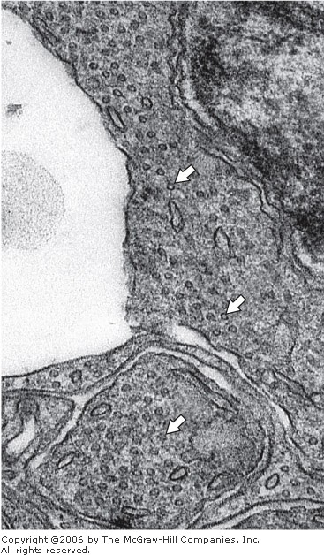


Figure 2-32

Electron micrograph of a section of a photosensitive retinal cell. Note the accumulation of transversely sectioned microtubules (arrows). Reduced slightly from x80,000.

Figure 2-33
Photomicrograph of the epithelium covering the inner surface of the respiratory airways. Most cells in this epithelium contain numerous cilia in their apices (free upper extremities). N, cell nuclei; M, cytoplasmic mucus secretion, which appears dark in this preparation. H&E stain. High magnification.


Figure 2-34

Schematic representation of microtubules, cilia, and centrioles. A: Microtubules as seen in the electron microscope after fixation with tannic acid in glutaraldehyde. The unstained tubulin subunits are delineated by the dense tannic acid. Cross sections of tubules reveal a ring of 13 subunits of dimers arranged in a spiral. Changes in microtubule length are due to the addition or loss of individual tubulin subunits. B: A cross section through a cilium reveals a core of microtubules called an axoneme. The axoneme consists of two central microtubules surrounded by nine microtubule doublets. In the doublets, microtubule A is complete and consists of 13 subunits, whereas microtubule B shares two or three heterodimers with A. When activated by ATP, the dynein arms link adjacent tubules and provide for the sliding of doublets against each other. C: Centrioles consist of nine microtubule triplets linked together in a pinwheel-like arrangement. In the triplets, microtubule A is complete and consists of 13 subunits, whereas microtubules B and C share tubulin subunits. Under normal circumstances, these organelles are found in pairs with the centrioles disposed at right angles to one another.

The subunit of a microtubule is a heterodimer composed of and tubulin molecules of closely related amino acid composition, each with a molecular mass of about 50 kDa.

Under appropriate conditions (in vivo or in vitro), tubulin subunits polymerize to form microtubules. With special staining procedures, tubulin can be seen as heterodimers organized into a spiral. A total of 13 units is present in one complete turn of the spiral (Figure 2–34).

Polymerization of tubulins to form microtubules in vivo is directed by a variety of structures collectively known as microtubule-organizing centers. These structures include cilia, basal bodies, and centrosomes. Microtubule growth, via subunit polymerization, occurs more rapidly at one end of existing microtubules. This end is referred to as the plus (+) end, and the other extremity is the minus (–) end. Tubulin polymerization is under control of the concentration of Ca2+ and of the microtubule-associated proteins, or MAPs. Microtubule stability is variable; for example, microtubules of cilia are stable, whereas microtubules of the mitotic spindle have a short duration. The antimitotic alkaloid colchicine binds specifically to tubulin, and when the complex tubulin–colchicine binds to microtubules, it prevents the addition of more tubulin in the plus (+) extremity. Mitotic microtubules are broken down because the depolymerization continues, mainly at the minus (–) end, and the lost tubulin units are not replaced. Another alkaloid that interferes with the mitotic microtubule is taxol, which accelerates the formation of microtubules but at the same time stabilizes them. All cytosolic tubulin is used in stable microtubules, and no tubulin is left for the formation of the mitotic spindle. Another alkaloid, vinblastine, acts by depolymerizing formed microtubules and, in a second step, aggregating to form paracrystalline arrays of tubulin.

Medical Application

The antimitotic alkaloids are useful tools in cell biology (eg, colchicine is used to arrest chromosomes in metaphase and to prepare karyotypes) and in cancer chemotherapy (eg, vinblastine, vincristine, and taxol are used to arrest cell proliferation in tumors). Because tumor cells proliferate rapidly, they are more affected by antimitotic drugs than are normal cells. However, chemotherapy has many undesirable consequences. For example, some normal blood-forming cells and the epithelial cells that cover the digestive tract also show a high rate of proliferation and are adversely affected by chemotherapy.


Cytoplasmic microtubules are stiff structures that play a significant role in the development and maintenance of cell shape. They are usually present in a proper orientation, either to effect development of a given cellular asymmetry or to maintain it. Procedures that disrupt microtubules result in the loss of this cellular asymmetry.

Microtubules also participate in the intracellular transport of organelles and vesicles. Examples include axoplasmic transport in neurons, melanin transport in pigment cells, chromosome movements by the mitotic spindle, and vesicle movements among different cell compartments. In each of these examples, movement is related to the presence of complex microtubule networks, and such activities are suspended if microtubules are disrupted. The transport guided by microtubules is under the control of special proteins called motor proteins, which use energy to move molecules and vesicles.

Microtubules provide the basis for several complex cytoplasmic components, including centrioles, basal bodies, cilia, and flagella. Centrioles are cylindrical structures (0.15 m in diameter and 0.3–0.5 m in length) composed primarily of short, highly organized microtubules (Figure 2–34). Each centriole shows nine sets of microtubules arranged in triplets. The microtubules are so close together that adjacent microtubules of a triplet share a common wall. Close to the nucleus of nondividing cells is a centrosome (Figure 2–35) made of a pair of centrioles surrounded by a granular material. In each pair, the long axes of the centrioles are at right angles to each other. Before cell division, more specifically during the S period of the interphase, each centrosome duplicates itself so that now each centrosome has two pairs of centrioles. During mitosis, the centrosomes divide in two, move to opposite poles of the cell, and become organizing centers for the microtubules of the mitotic spindle.

Figure 2-35
Drawing of a centrosome with its granular protein material surrounding a pair of centrioles, one shown at a right angle to the other. Each centriole is composed of nine bundles of microtubules, with three microtubules per bundle.

Cilia and flagella (singular, cilium, flagellum) are motile processes, covered by cell membrane, with a highly organized microtubule core. Ciliated cells typically possess a large number of cilia, each about 2–3 m in length. Flagellated cells have only one flagellum, with a length close to 100 m. In humans, the spermatozoa are the only cell type with a flagellum. The main function of cilia is to sweep fluid from the surface of cell sheets. Both cilia and flagella possess the same core organization.

This core consists of nine pairs of microtubules surrounding two central microtubules. This sheaf of microtubules, possessing a 9 + 2 pattern, is called an axoneme (Gr. axon, axis, + nema, thread). Each of the nine peripheral pairs shares a common wall (Figure 2–34). The microtubules in the central pair are enclosed within a central sheath. Adjacent peripheral pairs are linked to each other by protein bridges called nexins and to the central sheath by radial spokes. The microtubules of each pair are identified as A and B. Microtubule A is complete, with 13 heterodimers, whereas B has only 10 heterodimers (in a cross section). Extending from the surface of microtubule A are pairs of arms formed by the protein dynein, which has ATPase activity.

At the base of each cilium or flagellum is a basal body, essentially similar to a centriole, that controls the assembly of the axoneme.

Medical Application

Several mutations have been described in the proteins of the cilia and flagella. They are responsible for the immotile cilia syndrome, the symptoms of which are immotile spermatozoa, male infertility, and chronic respiratory infections caused by the lack of the cleansing action of cilia in the respiratory tract.

References
Afzelius BA, Eliasson R: Flagellar mutants in man: on the heterogeneity of the immotile-cilia syndrome. J Ultrastruct Res 1979;69:43. [PMID: 501788]
Aridor M, Balch WE: Integration of endoplasmic reticulum signaling in health and disease. Nat Med 1999;5:745. [PMID: 10395318]
Barrit GJ: Communication Within Animal Cells. Oxford University Press, 1992.
Becker WM et al: The World of the Cell, 4th ed. Benjamin/Cummings, 2000.
Bretscher MS: The molecules of the cell membrane. Sci Am 1985;253:100. [PMID: 2416050]
Brinkley BR: Microtubule organizing centers. Annu Rev Cell Biol 1985;1:145. [PMID: 3916316]
Brown MS et al: Recycling receptors: the round-trip itinerary of migrant membrane proteins. Cell 1983;32:663. [PMID: 6299572]
Cooper GM: The Cell: A Molecular Approach. ASM Press/Sinauer Associates, Inc., 1997.
DeDuve C: A Guided Tour of the Living Cell. Freeman, 1984.
DeDuve C: Microbodies in the living cell. Sci Am 1983;248:74.
Dustin P: Microtubules, 2nd ed. Springer-Verlag, 1984.
Farquhar MG: Progress in unraveling pathways of Golgi traffic. Annu Rev Cell Biol 1985;1:447. [PMID: 3916320]
Fawcett D: The Cell, 2nd ed. Saunders, 1981.
Krstíc RV: Ultrastructure of the Mammalian Cell. Springer-Verlag, 1979.
Mitchison TJ, Cramer LP: Actin-based cell motility and cell locomotion. Cell 1996;84:371. [PMID: 8608590]
Osborn M, Weber K: Intermediate filaments: cell-type-specific markers in differentiation and pathology. Cell 1982;31:303. [PMID: 6891619]
Pfeffer SR, Rothman JE: Biosynthetic protein transport and sorting in the endoplasmic reticulum. Annu Rev Biochem 1987;56:829. [PMID: 3304148]
Rothman J: The compartmental organization of the Golgi apparatus. Sci Am 1985;253:74. [PMID: 3929377]
Simons K, Ikonen E: How cells handle cholesterol. Science 2000;290:1721. [PMID: 11099405]
Tzagoloff A: Mitochondria. Plenum, 1982.
Weber K, Osborn M: The molecules of the cell matrix. Sci Am 1985;253:110. [PMID: 4071030]


Proteasomes, Peroxisomes, & Secretory Vesicles

Proteasomes

Proteasomes are multiple-protease complexes that digest proteins targeted for destruction by attachment to ubiquitin. Protein degradation is essential to remove excess enzyme and other proteins that become unnecessary to the cell after they perform their normal functions, and also to remove proteins that were incorrectly folded. Protein encoded by virus should also be destroyed. Proteasomes deal primarily with proteins as individual molecules, whereas lysosomes digest bulk material introduced into the cell or whole organelles and vesicles.

The proteasome has a core particle with the shape of a barrel made of four rings stacked on each other. At each end of the core particle is a regulatory particle that contains ATPase and recognizes proteins with ubiquitin molecules attached. Ubiquitin is a small protein (76 amino acids) found in all cells and is highly conserved during evolution—it has virtually the same structure from bacteria to humans. Ubiquitin targets proteins for destruction as follows. A molecule of ubiquitin binds to a lysine residue in the protein to be degraded. Then other ubiquitin molecules attach to the first one; the complex is recognized by the regulatory particle; the protein is unfolded by the ATPases using energy from ATP; and the protein is translocated into the core particle, where it is broken into peptides of about eight amino acids each. These peptides are transferred to the cytosol by a process yet unknown. The ubiquitin molecules are released by the regulatory particles for reuse.

The eight-amino acid peptides may be broken down to amino acids by cytosol enzymes, or they may have other destinations (eg, in some cells they participate in the immune response).

Peroxisomes (Microbodies)


Peroxisomes (peroxide + soma, body) are spherical membrane-limited organelles whose diameter ranges from 0.5 to 1.2 m (see Figure 2–39). Like the mitochondria, they utilize oxygen but do not produce ATP and do not participate directly in cellular metabolism. Peroxisomes oxidize specific organic substrates by removing hydrogen atoms that are transferred to molecular oxygen (O2). This activity produces hydrogen peroxide (H2O2), a substance that is very damaging to the cell. However, H2O2 is eliminated by the enzyme catalase, which is present in peroxisomes. Catalase transfers oxygen atoms from H2O2 to several compounds and also decomposes H2O2 to H2O and O2 (2 H2O2 2 H2O + O2). Catalase activity also has clinical implications. It degrades several toxic molecules and prescription drugs, particularly in liver and kidney peroxisomes. For example, 50% of ingested ethyl alcohol is degraded to acetic aldehyde in liver and kidney peroxisomes. Liver and kidney peroxisomes show a higher variation in their enzyme complement than do other peroxisomes. Their homogeneous matrix contains D- and L-amino oxidases, catalase, and hydroxyacid oxidase. In some species, but not humans, a crystalline nucleoid is present that is composed of urate oxidase.

Peroxisomes contain enzymes involved in lipid metabolism. Thus, the -oxidation of long-chain fatty acids (18 carbons and longer) is preferentially accomplished by peroxisomal enzymes that differ from their mitochondrial counterparts. Certain reactions leading to the formation of bile acids and cholesterol also have been localized in highly purified peroxisomal fractions.

Peroxisomal enzymes are synthesized on free cytosolic polyribosomes, with a small sequence of amino acids located near the carboxyl terminus that functions as an import signal. Proteins with this signal are recognized by receptors located in the membrane of peroxisomes and internalized into the organelle. The peroxisome grows in size and is divided into two smaller peroxisomes, by a mechanism not completely understood.

Medical Application

A large number of disorders arise from defective peroxisomal proteins, because this organelle is involved in several metabolic pathways. Probably the most common peroxisomal disorder is X-chromosome-linked adrenoleukodystrophy, caused by a defective integral membrane protein that participates in transporting very long-chain fatty acids into the peroxisome for -oxidation. Accumulation of these fatty acids in body fluids destroys the myelin sheaths in nerve tissue, causing severe neurological symptoms. Deficiency in peroxisomal enzymes causes the fatal Zellweger syndrome, with severe muscular impairment, liver and kidney lesions, and disorganization of the central and peripheral nervous systems. Electron microscopy reveals empty peroxisomes in liver and kidney cells of these patients.

Secretory Vesicles, or Granules

Secretory vesicles are found in those cells that store a product until its release is signaled by a metabolic, hormonal, or neural message (regulated secretion). These vesicles are surrounded by a membrane and contain a concentrated form of the secretory product (Figure 2–29). The contents of some secretory vesicles may be up to 200 times more concentrated than those in the cisternae of the RER. Secretory vesicles containing digestive enzymes are referred to as zymogen granules.

Figure 2-29
Electron micrograph of a pancreatic acinar cell from the rat. Numerous mature secretory granules (S) are seen in association with condensing vacuoles (C) and the Golgi complex (G). x18,900.

References
Afzelius BA, Eliasson R: Flagellar mutants in man: on the heterogeneity of the immotile-cilia syndrome. J Ultrastruct Res 1979;69:43. [PMID: 501788]
Aridor M, Balch WE: Integration of endoplasmic reticulum signaling in health and disease. Nat Med 1999;5:745. [PMID: 10395318]
Barrit GJ: Communication Within Animal Cells. Oxford University Press, 1992.
Becker WM et al: The World of the Cell, 4th ed. Benjamin/Cummings, 2000.
Bretscher MS: The molecules of the cell membrane. Sci Am 1985;253:100. [PMID: 2416050]
Brinkley BR: Microtubule organizing centers. Annu Rev Cell Biol 1985;1:145. [PMID: 3916316]
Brown MS et al: Recycling receptors: the round-trip itinerary of migrant membrane proteins. Cell 1983;32:663. [PMID: 6299572]
Cooper GM: The Cell: A Molecular Approach. ASM Press/Sinauer Associates, Inc., 1997.
DeDuve C: A Guided Tour of the Living Cell. Freeman, 1984.
DeDuve C: Microbodies in the living cell. Sci Am 1983;248:74.
Dustin P: Microtubules, 2nd ed. Springer-Verlag, 1984.
Farquhar MG: Progress in unraveling pathways of Golgi traffic. Annu Rev Cell Biol 1985;1:447. [PMID: 3916320]
Fawcett D: The Cell, 2nd ed. Saunders, 1981.
Krstíc RV: Ultrastructure of the Mammalian Cell. Springer-Verlag, 1979.
Mitchison TJ, Cramer LP: Actin-based cell motility and cell locomotion. Cell 1996;84:371. [PMID: 8608590]
Osborn M, Weber K: Intermediate filaments: cell-type-specific markers in differentiation and pathology. Cell 1982;31:303. [PMID: 6891619]
Pfeffer SR, Rothman JE: Biosynthetic protein transport and sorting in the endoplasmic reticulum. Annu Rev Biochem 1987;56:829. [PMID: 3304148]
Rothman J: The compartmental organization of the Golgi apparatus. Sci Am 1985;253:74. [PMID: 3929377]
Simons K, Ikonen E: How cells handle cholesterol. Science 2000;290:1721. [PMID: 11099405]
Tzagoloff A: Mitochondria. Plenum, 1982.
Weber K, Osborn M: The molecules of the cell matrix. Sci Am 1985;253:110. [PMID: 4071030]


Selasa, 21 Juni 2011

Lysosomes

Lysosomes are sites of intracellular digestion and turnover of cellular components. Lysosomes (Gr. lysis, solution, + soma, body) are membrane-limited vesicles that contain a large variety of hydrolytic enzymes (more than 40) whose main function is intracytoplasmic digestion (Figures 2–24, 2–25, and 2–26). Lysosomes are particularly abundant in cells exhibiting phagocytic activity (eg, macrophages, neutrophilic leukocytes). Although the nature and activity of lysosomal enzymes vary depending on the cell type, the most common enzymes are acid phosphatase, ribonuclease, deoxyribonuclease, proteases, sulfatases, lipases, and beta-glucuronidase. As can be seen from this list, lysosomal enzymes are capable of breaking down most biological macromolecules. Lysosomal enzymes have optimal activity at an acidic pH.

Figure 2-24
Photomicrograph of a kidney tubule whose lumen appears in the center as a long slit. The numerous dark-stained cytoplasmic granules are lysosomes (L), organelles abundant in these kidney cells. The cell nuclei (N), some showing a nucleolus, are also seen in the photograph as dark-stained corpuscles. Toluidine blue stain. High magnification.

Figure 2-25
Electron micrograph of a macrophage. Note the abundant cytoplasmic extensions (arrows). In the center is a centriole (C) surrounded by Golgi cisternae (G). Secondary lysosomes (L) are abundant. x15,000.

Figure 2-26
Electron micrograph showing four dark secondary lysosomes surrounded by numerous mitochondria.

Lysosomes, which are usually spherical, range in diameter from 0.05 to 0.5 micrometer and present a uniformly granular, electron-dense appearance in electron micrographs. In a few cells, such as macrophages and neutrophilic leukocytes, primary lysosomes are larger, up to 0.5 micrometer in diameter, and thus are just visible with the light microscope.

The enveloping membrane separates the lytic enzymes from the cytoplasm, preventing the lysosomal enzymes from attacking and digesting cytoplasmic components. The fact that the lysosomal enzymes are practically inactive at the pH of the cytosol (~7.2) is an additional protection of the cell against leakage of lysosomal enzymes.

Lysosomal enzymes are synthesized and segregated in the RER and subsequently transferred to the Golgi complex, where the enzymes are modified and packaged as lysosomes. These enzymes have oligosaccharides attached to them with one or more of the mannose residues phosphorylated at the 6´ position by a phosphotransferase. There are receptors for mannose 6-phosphate-containing proteins in the RER and Golgi complex that allow these proteins to be diverted from the main secretory pathway and segregated in lysosomes.

Lysosomes that have not entered into a digestive event are identified as primary lysosomes.

Lysosomes can digest material taken into the cell from its environment. The material is taken into a phagosome or phagocytic vacuole (Figure 2–27); primary lysosomes then fuse with the membrane of the phagosome and empty their hydrolytic enzymes into the vacuole. Digestion follows, and the composite structure is now termed a secondary lysosome.

Figure 2-27
Current concepts of the functions of lysosomes. Synthesis occurs in the rough endoplasmic reticulum (RER), and the enzymes are packaged in the Golgi complex. Note the heterophagosomes, in which bacteria are being destroyed, and the autophagosomes, with RER and mitochondria in the process of digestion. Heterophagosomes and autophagosomes are secondary lysosomes. The result of their digestion can be excreted, but sometimes the secondary lysosome creates a residual body, containing remnants of undigested molecules. In some cells, such as osteoclasts, the lysosomal enzymes are secreted to the extracellular environment. Nu, nucleolus.

Secondary lysosomes are generally 0.2–2 micrometer in diameter and present a heterogeneous appearance in electron microscopes because of the wide variety of materials they may be digesting.

After digestion of the contents of the secondary lysosome, nutrients diffuse through the lysosomal-limiting membrane and enter the cytosol. Indigestible compounds are retained within the vacuoles, which are now called residual bodies (Figures 2–27 and 2–28). In some long-lived cells (eg, neurons, heart muscle), large quantities of residual bodies accumulate and are referred to as lipofuscin, or age pigment.

Figure 2-28
Section of a pancreatic acinar cell showing autophagosomes. Upper right: Two portions of the rough endoplasmic reticulum segregated by a membrane. Center: An autophagosome containing mitochondria (arrow) plus rough endoplasmic reticulum. Left: A residual body, with indigestible material. Arrowhead shows a cluster of coated vesicles.

Another function of lysosomes concerns the turnover of cytoplasmic organelles. Under certain conditions, a membrane may enclose organelles or portions of cytoplasm. Primary lysosomes fuse with this structure and initiate the lysis of the enclosed cytoplasm. The resulting secondary lysosomes are known as autophagosomes (Gr. autos, self, + phagein, to eat, + soma, body), indicating that their contents are intracellular in origin. Cytoplasmic digestion by autophagosomes is enhanced in secretory cells that have accumulated excess secretory product. The digested products of lysosomal hydrolysis are recycled by the cell to be reutilized by the cytoplasm.

Medical Apllication
In some cases, primary lysosomes release their contents extracellularly, and their enzymes act in the extracellular milieu. An example is the destruction of bone matrix by the collagenases synthesized and released by osteoclasts during normal bone tissue formation (see Chapter 8: Bone). Lysosomal enzymes acting in the extracellular milieu also play a significant role in the response to inflammation or injury. Several possible pathways relating to lysosome activities are schematically illustrated in Figure 2–27.

Lysosomes play an important role in the metabolism of several substances in the human body, and consequently many diseases have been ascribed to deficiencies of lysosomal enzymes. In metachromatic leukodystrophy, there is an intracellular accumulation of sulfated cerebrosides caused by lack of lysosomal sulfatases. In most of these diseases, a specific lysosomal enzyme is absent or inactive, and certain molecules (eg, glycogen, cerebrosides, gangliosides, sphingomyelin, glycosaminoglycans) are not digested. As a result, these substances accumulate in the cells, interfering with their normal functions. This diversity of affected cell types explains the variety of clinical symptoms observed in lysosomal diseases (Table 2–3).

Table 2–3. Examples of Diseases Caused by Lysosomal Enzyme Failure and Accumulation of Undigested Material in Different Cell Types.
Disease Faulty Enzyme Main Organs Affected
Hurler disease alfa-L-Iduronidase  Skeleton and nervous system
Sanfilippo syndrome A Heparan sulfate sulfamidase Skeleton and nervous system
Tay-Sachs Hexosaminidase-A Nervous system
Gaucher beta-D-Glycosidase  Liver and spleen
I-cell disease Phosphotransferase Skeleton and nervous system

I-cell disease (inclusion cell disease) is a rare inherited condition clinically characterized by defective physical growth and mental retardation and is due to a deficiency in a phosphorylating enzyme normally present in the Golgi complex. Lysosomal enzymes coming from the RER are not phosphorylated in the Golgi complex. Nonphosphorylated protein molecules are not separated to form lysosomes, instead following the main secretory pathway. The secreted lysosomal enzymes are present in the blood of patients with I-cell disease, whereas their lysosomes are empty. Cells of these patients show large inclusion granules that interfere with normal cellular metabolism.

References
Afzelius BA, Eliasson R: Flagellar mutants in man: on the heterogeneity of the immotile-cilia syndrome. J Ultrastruct Res 1979;69:43. [PMID: 501788]
Aridor M, Balch WE: Integration of endoplasmic reticulum signaling in health and disease. Nat Med 1999;5:745. [PMID: 10395318]
Barrit GJ: Communication Within Animal Cells. Oxford University Press, 1992.
Becker WM et al: The World of the Cell, 4th ed. Benjamin/Cummings, 2000.
Bretscher MS: The molecules of the cell membrane. Sci Am 1985;253:100. [PMID: 2416050]
Brinkley BR: Microtubule organizing centers. Annu Rev Cell Biol 1985;1:145. [PMID: 3916316]
Brown MS et al: Recycling receptors: the round-trip itinerary of migrant membrane proteins. Cell 1983;32:663. [PMID: 6299572]
Cooper GM: The Cell: A Molecular Approach. ASM Press/Sinauer Associates, Inc., 1997.
DeDuve C: A Guided Tour of the Living Cell. Freeman, 1984.
DeDuve C: Microbodies in the living cell. Sci Am 1983;248:74.
Dustin P: Microtubules, 2nd ed. Springer-Verlag, 1984.
Farquhar MG: Progress in unraveling pathways of Golgi traffic. Annu Rev Cell Biol 1985;1:447. [PMID: 3916320]
Fawcett D: The Cell, 2nd ed. Saunders, 1981.
Krstíc RV: Ultrastructure of the Mammalian Cell. Springer-Verlag, 1979.
Mitchison TJ, Cramer LP: Actin-based cell motility and cell locomotion. Cell 1996;84:371. [PMID: 8608590]
Osborn M, Weber K: Intermediate filaments: cell-type-specific markers in differentiation and pathology. Cell 1982;31:303. [PMID: 6891619]
Pfeffer SR, Rothman JE: Biosynthetic protein transport and sorting in the endoplasmic reticulum. Annu Rev Biochem 1987;56:829. [PMID: 3304148]
Rothman J: The compartmental organization of the Golgi apparatus. Sci Am 1985;253:74. [PMID: 3929377]
Simons K, Ikonen E: How cells handle cholesterol. Science 2000;290:1721. [PMID: 11099405]
Tzagoloff A: Mitochondria. Plenum, 1982.
Weber K, Osborn M: The molecules of the cell matrix. Sci Am 1985;253:110. [PMID: 4071030]

Golgi Complex (Golgi Apparatus)

The Golgi complex completes posttranslational modifications and packages and places an address on products that have been synthesized by the cell. This organelle is composed of smooth membrane-limited cisternae (Figures 2–21, 2–22, and 2–23). In highly polarized cells, such as mucus-secreting goblet cells (Figure 4–31), the Golgi complex occupies a characteristic position in the cytoplasm between the nucleus and the apical plasma membrane.

Figure 2-21
Three-dimensional representation of a Golgi complex. Through transport vesicles that fuse with the Golgi cis face, the complex receives several types of molecules produced in the rough endoplasmic reticulum (RER). After Golgi processing, these molecules are released from the Golgi trans face in larger vesicles to constitute secretory vesicles, lysosomes, or other cytoplasmic components.


Figure 2-22
Electron micrograph of a Golgi complex of a mucous cell. To the right is a cisterna (arrow) of the rough endoplasmic reticulum containing granular material. Close to it are small vesicles containing this material. This is the cis face of the complex. In the center are flattened and stacked cisternae of the Golgi complex. Dilatations can be observed extending from the ends of the cisternae. These dilatations gradually detach themselves from the cisternae and fuse, forming the secretory granules (1, 2, and 3). This is the trans face. Near the plasma membrane of two neighboring cells is endoplasmic reticulum with a smooth section (SER) and a rough section (RER). x30,000. Inset: The Golgi complex as seen in 1-m sections of epididymis cells impregnated with silver. x1200.

Figure 2-23
Main events occurring during trafficking and sorting of proteins through the Golgi complex. Numbered at the left are the main molecular processes that take place in the compartments indicated. Note that the labeling of lysosomal enzymes starts early in the cis Golgi network. In the trans Golgi network, the glycoproteins combine with specific receptors that guide them to their destination. On the left side of the drawing is the returning flux of membrane, from the Golgi to the endoplasmic reticulum. (Redrawn and reproduced, with permission, from Junqueira LC, Carneiro J: Biologia Celular e Molecular, 6th ed. Editora Guanabara, 1997.)

In most cells, there is also polarity in Golgi structure and function. Near the Golgi complex, the RER can sometimes be seen budding off small vesicles (transport vesicles) that shuttle newly synthesized proteins to the Golgi complex for further processing. The Golgi cisterna nearest this point is called the forming, convex, or cis face. On the opposite side of the Golgi complex, which is the maturing, concave, or trans face, large Golgi vacuoles accumulate (Figure 2–21). These are sometimes called condensing vacuoles.
These structures bud from the Golgi cisternae, generating vesicles that will transport proteins to various sites. Cytochemical methods and the electron microscope have shown that the Golgi cisternae present different enzymes at different cis–trans levels and that the Golgi complex is important in the glycosylation, sulfating, phosphorylation, and limited proteolysis of proteins. Furthermore, the Golgi complex initiates packing, concentration, and storage of secretory products. Figure 2–23 provides an overall view of the currently accepted concepts regarding transit of material through the Golgi complex.

References
Afzelius BA, Eliasson R: Flagellar mutants in man: on the heterogeneity of the immotile-cilia syndrome. J Ultrastruct Res 1979;69:43. [PMID: 501788]
Aridor M, Balch WE: Integration of endoplasmic reticulum signaling in health and disease. Nat Med 1999;5:745. [PMID: 10395318]
Barrit GJ: Communication Within Animal Cells. Oxford University Press, 1992.
Becker WM et al: The World of the Cell, 4th ed. Benjamin/Cummings, 2000.
Bretscher MS: The molecules of the cell membrane. Sci Am 1985;253:100. [PMID: 2416050]
Brinkley BR: Microtubule organizing centers. Annu Rev Cell Biol 1985;1:145. [PMID: 3916316]
Brown MS et al: Recycling receptors: the round-trip itinerary of migrant membrane proteins. Cell 1983;32:663. [PMID: 6299572]
Cooper GM: The Cell: A Molecular Approach. ASM Press/Sinauer Associates, Inc., 1997.
DeDuve C: A Guided Tour of the Living Cell. Freeman, 1984.
DeDuve C: Microbodies in the living cell. Sci Am 1983;248:74.
Dustin P: Microtubules, 2nd ed. Springer-Verlag, 1984.
Farquhar MG: Progress in unraveling pathways of Golgi traffic. Annu Rev Cell Biol 1985;1:447. [PMID: 3916320]
Fawcett D: The Cell, 2nd ed. Saunders, 1981.
Krstíc RV: Ultrastructure of the Mammalian Cell. Springer-Verlag, 1979.
Mitchison TJ, Cramer LP: Actin-based cell motility and cell locomotion. Cell 1996;84:371. [PMID: 8608590]
Osborn M, Weber K: Intermediate filaments: cell-type-specific markers in differentiation and pathology. Cell 1982;31:303. [PMID: 6891619]
Pfeffer SR, Rothman JE: Biosynthetic protein transport and sorting in the endoplasmic reticulum. Annu Rev Biochem 1987;56:829. [PMID: 3304148]
Rothman J: The compartmental organization of the Golgi apparatus. Sci Am 1985;253:74. [PMID: 3929377]
Simons K, Ikonen E: How cells handle cholesterol. Science 2000;290:1721. [PMID: 11099405]
Tzagoloff A: Mitochondria. Plenum, 1982.
Weber K, Osborn M: The molecules of the cell matrix. Sci Am 1985;253:110. [PMID: 4071030]


Postingan Lebih Baru Postingan Lama Beranda
Your 1:1 Traffic Exchange


Chat Box


Top Commentators

Give Me +1

Admin

Foto Saya
I created this blog just to support the activities of my learning about histology and others

Followers

Rank

 
Copyright © 2011 De Histology

Templates by Poncol Gate | CSS3 by David Walsh | Powered by {N}Code & Blogger