Cell proliferation for renewal and growth is a process of self-evident physiological significance. Less evident, but no less important for body functions and health, is the process of programmed cell death called apoptosis. A few examples of apoptosis will illustrate its significance.
Most T lymphocytes originating in the thymus have the ability to attack and destroy body components and would cause serious damage if they entered the blood circulation. Inside the thymus, these T lymphocytes receive signals that activate the apoptotic program encoded in their chromosomes. They are destroyed by apoptosis before leaving the thymus (see Chapter 14: Lymphoid Organs).
Medical Application
Most cells of the body can activate their apoptotic program when major changes occur in their DNA for example, just before a tumor appears, when a number of mutations have already accumulated in the DNA. In this way, apoptosis prevents the proliferation of malignant cells that develop as a result of accumulated mutations in the DNA. To form a clone and develop into a tumor, the malignant cell needs to deactivate the genes that control the apoptotic process.
Apoptosis was first discovered in developing embryos, where programmed cell death is an essential process for shaping the embryo (morphogenesis). Later investigators observed that apoptosis is also a common event in the tissues of normal adults.
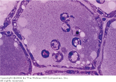
In apoptosis, the cell and its nucleus become compact, decreasing in size. At this stage the apoptotic cell shows a dark-stained nucleus (pyknotic nucleus), easily identified with the light microscope (Figure 3-24). Next, the chromatin is cut into pieces by DNA endonucleases. During apoptosis the cell shows cytoplasmic large vesicles (blebs) that detach from the cell surface (Figure 3-25). These detached fragments are contained within the plasma membrane, which is changed in such a way that all cell remnants are readily engulfed, or phagocytosed, mainly by macrophages. However, in macrophages the apoptotic fragments do not elicit the synthesis of the molecules that triggers the inflammatory process (see below).
Figure 3-24
Section of a mammary gland from an animal whose lactation was interrupted for 5 days. Note atrophy of the epithelial cells and dilation of the alveolar lumen, which contains several detached cells in the process of apoptosis, as seen from the nuclear alterations. PT stain. Medium magnification.
Figure 3-25
Electron micrograph of a cell in apoptosis showing that its cytoplasm is undergoing a process of fragmentation in blebs that preserve their plasma membranes. These blebs are phagocytosed by macrophages without eliciting an inflammatory reaction. No cytoplasmic substances are released into the extracellular space.
| References
|


0 comments:
Posting Komentar